

Mean Platelet Volume is not a number, it is a hypothesis about the bone marrow. Pull an MPV from a complete blood count, and you are really deciding whether the platelets on the smear are old and exhausted (low MPV, suppressed marrow) or young and reactive (high MPV, peripheral destruction with a marrow that is keeping up). The number sits beside the platelet count, the smear review, and the clinical context, and the differential diagnosis changes entirely depending on which of those three jobs the MPV is doing. A clinician who treats MPV as a single threshold misses most of the signal; a clinician who pairs it with the platelet count and the analyzer method catches the ITP from the reactive thrombocytosis every time.
What MPV Measures and Why It Varies
MPV is the mean size of platelets reported by an automated hematology analyzer, expressed in femtolitres (fL). It is the geometric companion to the platelet count, which counts the number. Platelets are anucleate fragments of megakaryocytes, and their size is set by the megakaryocyte’s ploidy at the moment of shedding. A megakaryocyte that responds to a high platelet demand makes larger, more granular platelets; a megakaryocyte that has been suppressed by chemotherapy or aplasia makes small, older platelets. That is the entire physiology: MPV is a real-time readout of whether the megakaryocyte is currently stressed.
The reference range is approximately 7.5 to 11.5 fL in most analyzers, but the range is method-dependent. Impedance counters (Coulter principle) tend to read 0.5 to 1.0 fL higher than optical counters; immunofluorescence on a fluorescence flow channel (PLT-F on Sysmex XN) tracks platelet volume closer to the true mean and is the reference to compare against. Comparing an MPV from a Sysmex XN against one from a Beckman Coulter DxH is not a fair comparison; it is two different measurements of the same number.
A second pre-analytical issue: platelets swell in EDTA over the first two hours after collection. A specimen analyzed at 30 minutes will read 0.3 to 0.8 fL lower than the same specimen at two hours. Most modern analyzers normalize this with a time-stamp, but older labs and some point-of-care devices do not. Compare within the same lab, on the same analyzer, at a consistent time after draw.
For a deeper exploration of MPV interpretation, the MPV Analyzer tool applies this exact framework, including the MPV/platelet-count pairing.
The Reference Range and Where It Comes From
The 7.5 to 11.5 fL range is not a hard biological constant. It is a 95 percent interval derived from healthy donor cohorts in the late 1980s and early 1990s (Bath 1996, followed by Vagdatli 2010), and the cohort’s demographics, analyzer mix, and anticoagulant choice all shape the number. Modern Sysmex XN-series normal cohorts sit closer to 9.0 to 12.5 fL; older Coulter STKS cohorts sit closer to 7.0 to 10.5 fL. Labs that report a single universal reference interval without method-specific calibration are reporting a fiction.
Three reference points to keep in mind:
- Lower bound (≈ 7.5 fL): below 7.0 fL on a calibrated analyzer is small old platelets, almost always from suppressed marrow production. Iron deficiency can sit here during the regenerative phase, when the marrow is producing small, poorly granulated cells. Wiskott-Aldrich syndrome has very small platelets by definition (WAS-platelet volume is a diagnostic feature, not an interpretation).
- Upper bound (≈ 11.5 fL): above 12.0 fL on a calibrated analyzer is large young platelets. In a healthy outpatient this is rare; in a recovering sepsis patient, a post-MI patient, or a patient with immune thrombocytopenia (ITP), it is the expected pattern.
- Method mismatch: a number that looks high on a Coulter may look normal on a Sysmex. Trust the analyzer’s own reference, not the textbook.
The reference range is a starting hypothesis, not a diagnosis. Use the MPV Analyzer tool to enter your lab’s exact number, see where it sits in the calibrated range, and to bring the platelet count in for the refined interpretation.
The MPV / Platelet Count Pairing Matrix
The single most useful thing a clinician can do with an MPV is pair it with the platelet count. The pairing splits the differential into four quadrants, and each quadrant points to a different category of disease.
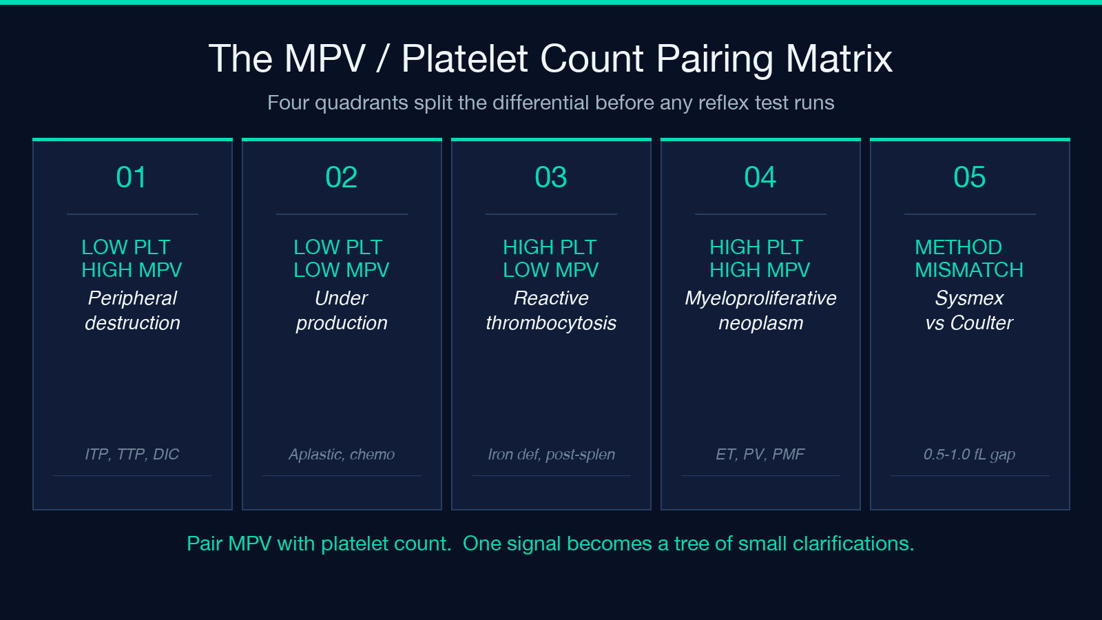
Low platelets + high MPV (low count, big young platelets): peripheral destruction with a marrow that is keeping up. The classic example is immune thrombocytopenia (ITP). The marrow sees platelet loss and ramps up megakaryocyte ploidy, releasing large young platelets. Disseminated intravascular coagulation (DIC) and thrombotic thrombocytopenic purpura (TTP) sit here too, but with other markers (D-dimer, schistocytes, haptoglobin) that dominate the picture.
Low platelets + low MPV (low count, small old platelets): underproduction. Aplastic anemia, recent chemotherapy or radiation, recent platelet transfusion (where most of the circulating platelets are donor-derived and older), and Wiskott-Aldrich syndrome all live here. This is the quadrant where the marrow is the problem.
High platelets + low MPV (high count, small platelets): reactive thrombocytosis. The marrow is producing more platelets, but they are small because the trigger is not stress-driven. Iron deficiency, post-splenectomy state, chronic inflammation, and malignancy-associated thrombocytosis fall here. Essential thrombocythemia can mimic it closely and is the differential that matters most.
High platelets + high MPV (high count, big platelets): myeloproliferative neoplasm territory. Essential thrombocythemia, polycythemia vera with thrombocytosis, primary myelofibrosis early phase, and chronic myeloid leukemia in some presentations. This quadrant warrants a JAK2 V617F or CALR reflex test before anything else.
Pairing MPV with the platelet count turns one signal into a tree of small-leaf clarifications. The pairing is the entire diagnostic value of the test.
What a Low MPV Tells You
A low MPV (under 7.5 fL, method-dependent) almost always points to suppressed marrow production. The marrow is not stressed, or it is being suppressed from outside (drugs, iron deficiency, marrow infiltration), or it has been physically damaged (radiation, chemotherapy, aplastic anemia). Small platelets are old platelets — they have circulated long enough to lose volume, and a marrow that is making them is not making fresh ones.
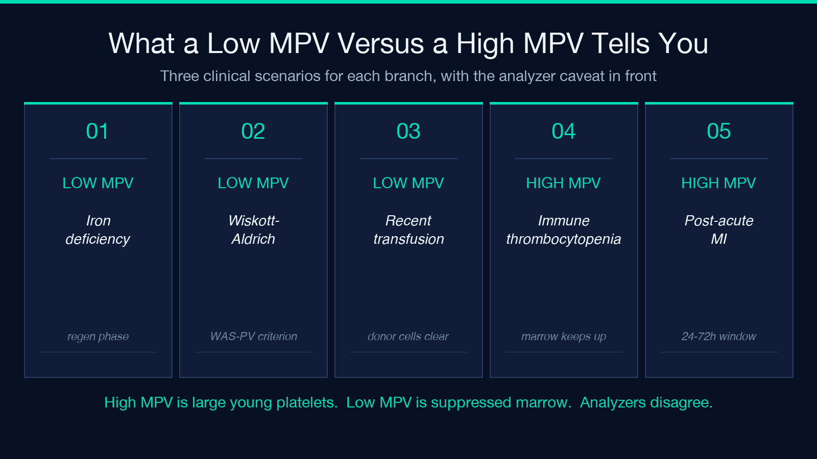
Three clinical scenarios where low MPV is the lead finding:
- Iron deficiency anemia in the regenerative phase: during iron repletion, the marrow ramps up production but the cells are still small because iron is the rate-limiting step. MPV is low because the platelets being released are still iron-poor. This is the inverse of the usual iron deficiency pattern (where everything else is small) and resolves over weeks of iron.
- Wiskott-Aldrich syndrome: a hereditary disorder with characteristically small platelets. MPV is part of the diagnostic criteria, not just an interpretation.
- Recent platelet transfusion: donor platelets are stored and smaller on the analyzer. MPV in a recently transfused patient is meaningless until the donor platelets clear, typically 3 to 5 days.
The MPV Analyzer tool handles the platelet-count pairing in the low-MPV branch automatically, including the iron-regeneration distinction and the post-transfusion caveat.
What a High MPV Tells You
A high MPV (above 11.5 fL, method-dependent) is large young platelets — a marrow that is stressed and pushing out fresh, large cells. In most patients this is reactive, but in some it is the lead finding for a primary marrow disorder.
Three clinical scenarios where high MPV is the lead finding:
- Immune thrombocytopenia (ITP): the marrow is compensating for peripheral destruction. Large young platelets are the rule, and MPV can climb above 12.5 fL in active disease. Pairing with the platelet count (which will be low) confirms the pattern.
- Post-splenectomy state: the spleen normally sequesters older platelets; without it, the circulating pool skews young. MPV stays mildly elevated for life.
- Post-acute myocardial infarction: platelets activated in the infarct zone release larger, more reactive platelets, and a high MPV 24 to 72 hours after an MI is a marker of adverse prognosis. This is one of the few places where MPV alone, without the pairing, has independent prognostic value.
The high-MPV branch is where the analyzer’s calibration matters most. A Sysmex XN with PLT-F will read MPV 0.5 to 1.0 fL lower than a Beckman Coulter DxH on the same specimen, so a “high” MPV on a Sysmex XN is not necessarily “high” on a Beckman Coulter.
The EDTA Pre-Analytical Caveat
Platelets swell in EDTA over time. A specimen analyzed at 30 minutes post-draw will read 0.3 to 0.8 fL lower than the same specimen at two hours. This is a real, well-characterized effect (Vagdatli 2010), and it has a clinical consequence: a patient whose CBC is repeated on the same specimen at two different labs may get two different MPV reports, and the difference is not error — it is biology.
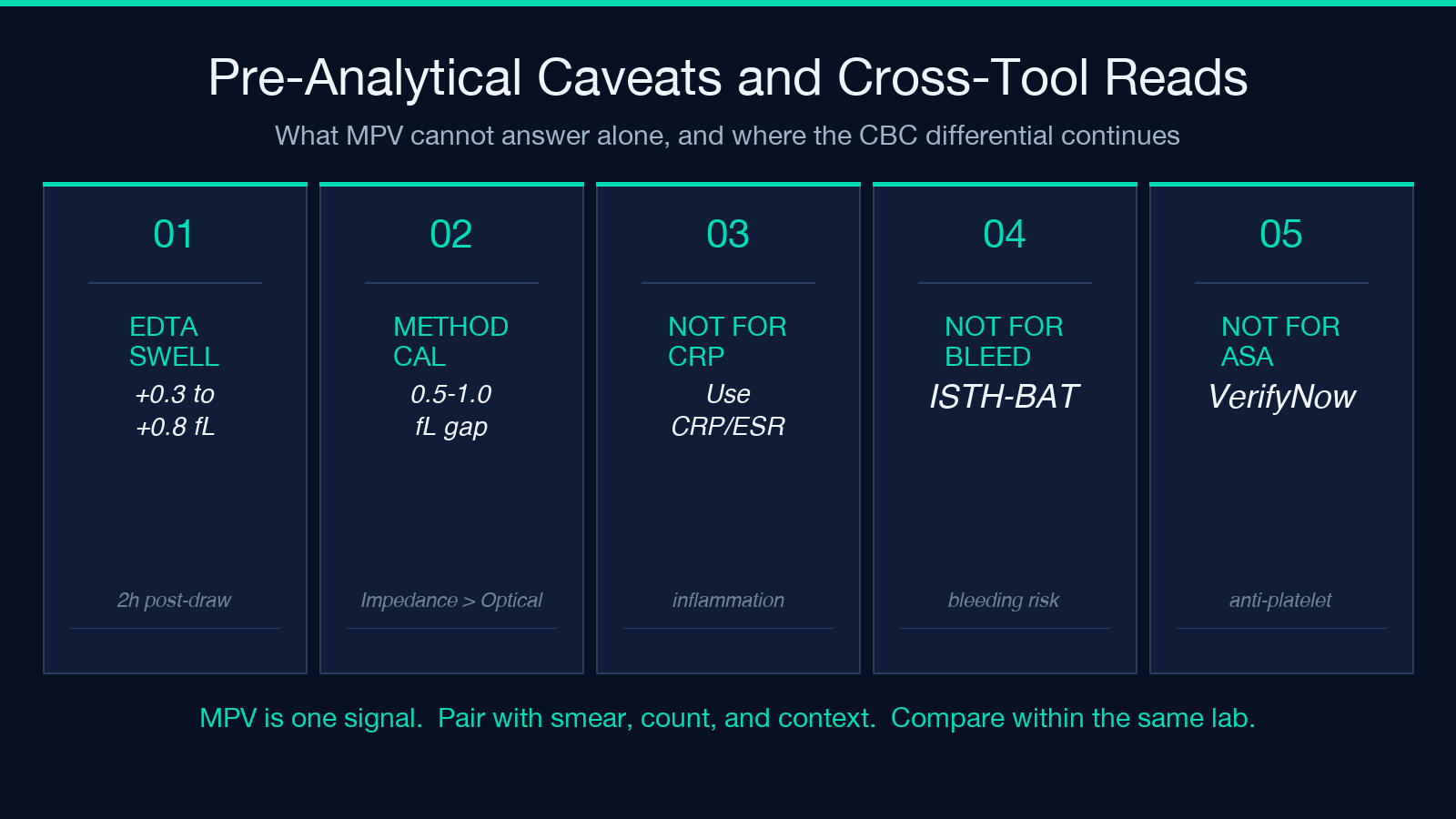
Two practical rules:
- Compare within the same lab. If a patient has serial MPVs from two different analyzers, treat the trend as informational, not diagnostic. The MPV trajectory is real, but the absolute number is not portable.
- If the MPV is borderline and the clinical question is sharp, request a citrate tube (blue top) repeat. Citrate does not cause platelet swelling, and the resulting MPV is closer to the in-vivo value.
For pediatric clinicians, the Absolute Neutrophil Count Calculator handles a related CBC-pair interpretation with the same pre-analytical discipline.
When MPV Alone Is Not Enough
MPV is a useful first-pass signal, but it has three known blind spots that no amount of calibration fixes:
- Acute inflammation: CRP and ESR rise faster and more reliably than MPV. Use MPV to answer the platelet-kinetics question, not the inflammation question.
- Bleeding risk: MPV bleeding does not predict bleeding risk in isolation. Use a bleeding-score tool (ISTH-BAT, WHO bleeding scale) for the bleeding question.
- Antiplatelet therapy response: MPV is not a validated monitor for aspirin or clopidogrel response. Platelet-function testing (VerifyNow, light transmission aggregometry) is the appropriate tool for that question.
MPV is one signal in a multi-signal dashboard. Pair it with the platelet count, the smear review, and the clinical context, and you have the differential. Try the MPV Analyzer tool for the pairing logic, the EDTA caveat, and the calibrated reference range.
Cross-Tool Reading List
For clinicians who want to extend MPV interpretation into the rest of the CBC differential:
– Reticulocyte Production Index Calculator — pairs MPV with marrow production rate. – Red Cell Distribution Width Analyzer — RDW is the size analog for red cells; the framework translates.
For broader CBC interpretation, the MPV Analyzer tool is the entry point. Explore more tools at elysiatools.com.
