
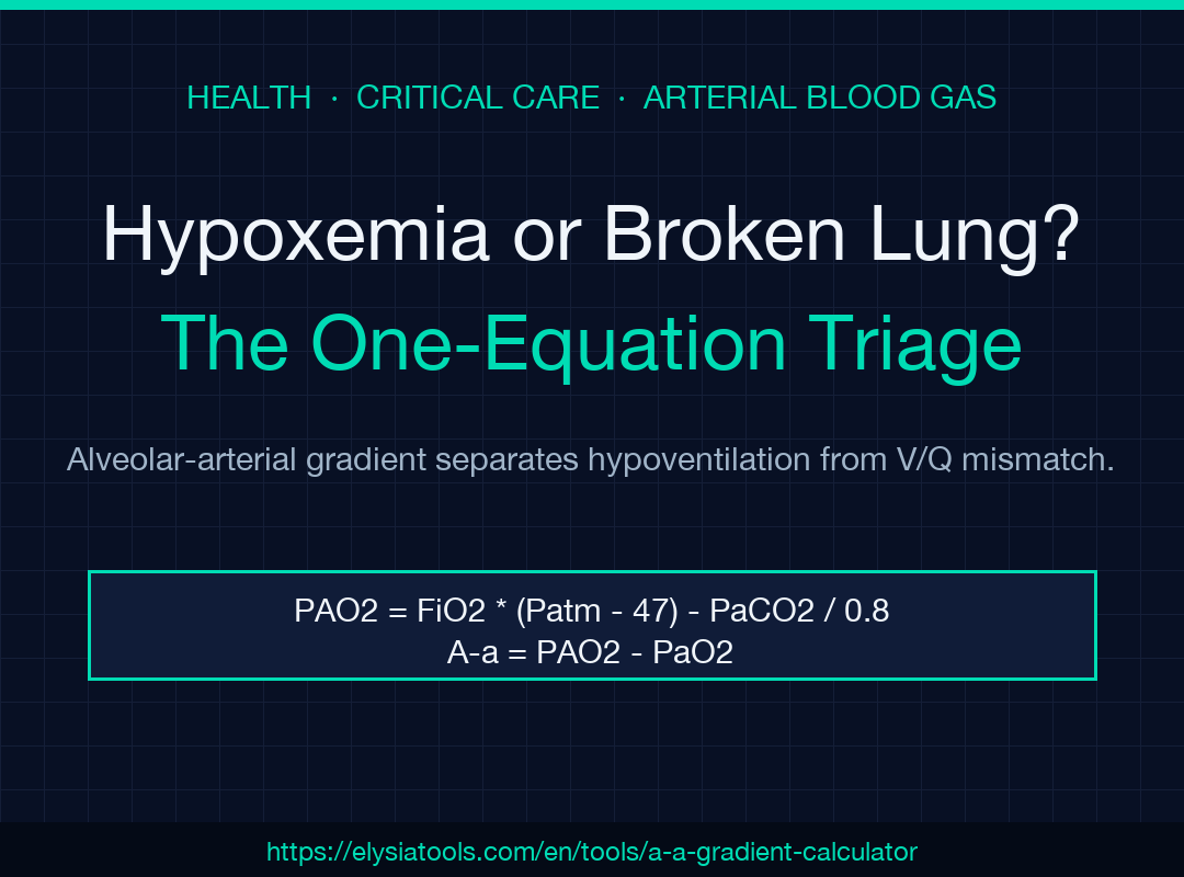
> Lead phrase. When the pulse oximeter reads 88% and the ABG comes back, every clinician at the bedside eventually asks the same question: is this hypoventilation, or is something in the lung actually broken? The A-a gradient — the difference between alveolar and arterial PO₂ — answers that in one equation.
The alveolar gas equation predicts how much oxygen should be in the alveolus given the FiO₂ and the barometric pressure, and arterial PaO₂ measures how much actually crossed into the blood. A normal gap means the lung is transferring oxygen as expected, so hypoxemia must be coming from somewhere else (low ventilation, low inspired oxygen, or low atmospheric pressure at altitude). A widened gap means there is a real intrapulmonary problem — V/Q mismatch, shunt, diffusion block, or low mixed-venous O₂. The A-a Gradient calculator runs the math and labels the result against the age-adjusted normal so the answer shows up before the next set of vitals does.
Why the A-a Gradient Matters at the Bedside
The ABG prints three numbers an intern actually needs: PaO₂, PaCO₂, FiO₂. Alone, they tell you the patient is hypoxemic but not why. The A-a gradient closes the loop. If the gap is normal, the clinician can skip a pulmonary workup, look at the drive to breathe (opioids, neuromuscular weakness, severe metabolic alkalosis), and treat accordingly. If the gap is widened, the differential narrows to V/Q mismatch (pneumonia, COPD, asthma, PE, pulmonary edema), shunt (ARDS, hepatopulmonary syndrome, intracardiac right-to-left), diffusion limitation (interstitial lung disease), or low mixed-venous O₂ (sepsis, shock, low cardiac output). One number, four destinations.
The Alveolar Gas Equation Walkthrough
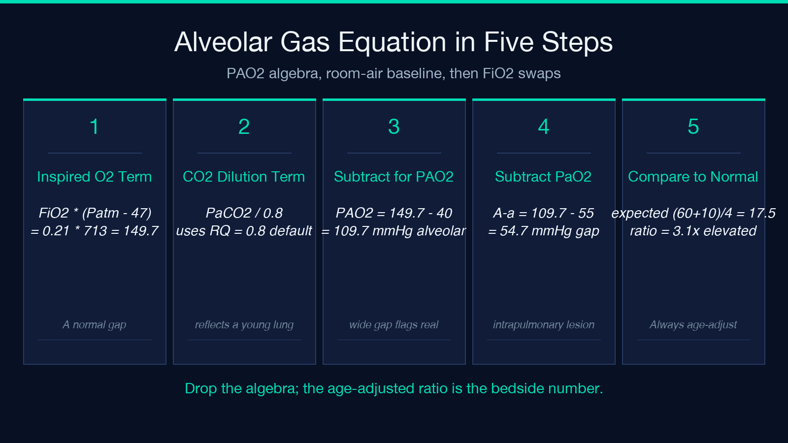
The math behind the gradient is short enough to do at the whiteboard.
> PAO₂ = FiO₂ × (P_atm − 47) − PaCO₂ / 0.8
The first term is the inspired oxygen partial pressure: 21% of dry barometric pressure at sea level, 47 mmHg subtracted for the water vapor pressure in the trachea. The second term is the alveolar dilution by CO₂ — the respiratory quotient (default 0.8) assumes the patient is burning a mixed diet of carbohydrate, fat, and protein. Lower the barometric pressure and PAO₂ drops. Lower the PaCO₂ and PAO₂ rises (hyperventilation packs more oxygen into each alveolar space). Lower the FiO₂ and PAO₂ falls in proportion.
Subtracting the measured PaO₂ gives the gradient:
> A-a = PAO₂ − PaO₂
The Age-Adjusted Normal Range
A “normal” A-a gradient is not 0 — small mismatches always exist between alveolar and arterial sides, and the gap widens with age. The canonical teaching rule is:
> Normal A-a ≈ (Age + 10) / 4
A 20-year-old patient breathing room air should have a gradient under about 7-8 mmHg. A 60-year-old can run up to ~17 mmHg and still be physiologically normal. A 90-year-old can hit 25 mmHg. The age cutoff matters because the alternative — a single fixed threshold like “<15 mmHg” — produces false positives on every elderly patient and false negatives on every teenager. The calculator uses (Age + 10) / 4 as its denominator and reports the gradient as a multiple of normal (e.g. “3.1× expected normal”), which keeps the interpretation scale-invariant across decades.
The Hypoxemia Decision Tree
Once the gradient is computed, the bedside logic follows two branches.
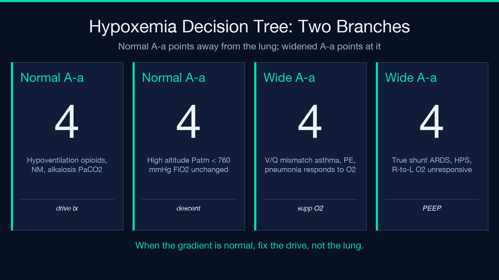
Normal gradient. Hypoxemia is not in the lung. Think hypoventilation (PaCO₂ > 45 mmHg suggests opioid, neuromuscular weakness, severe CNS depression); think high altitude (P_atm < 760 mmHg — Denver, Leadville, Mexico City); think low inspired oxygen (rebreather mask malfunction, hypoxic mix on a ventilator, sealed environment). Treat by correcting the drive (naloxone, BiPAP, ventilatory support) or the FiO₂ (supplemental oxygen, descent from altitude).
Widened gradient. Hypoxemia is in the lung. The next move is the response to supplemental oxygen. If PaO₂ jumps when FiO₂ goes from 0.21 to 1.0, the cause is V/Q mismatch — pneumonia, asthma, COPD exacerbation, pulmonary edema, early PE. If PaO₂ barely budges, the cause is true shunt — ARDS, hepatopulmonary syndrome, complete airway obstruction, large right-to-left intracardiac shunt. Diffusion limitation (interstitial fibrosis, ILD) sits in between — usually responds to high FiO₂ but never normalizes. Low mixed-venous O₂ (sepsis, cardiac arrest, severe anemia) shows up as a widened gradient too, because the deoxygenated blood returning to the lung cannot catch up no matter how good the alveolar side is.
The Worked Examples
Two ABGs illustrate the wide range of conclusions the gradient drives.
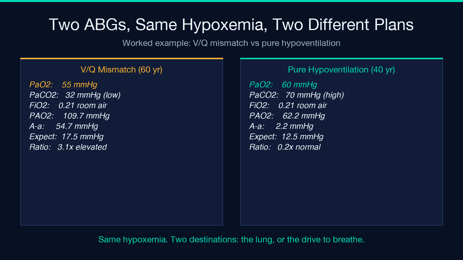
Example 1 — elevated A-a, V/Q mismatch. A 60-year-old patient on room air with PaO₂ 55, PaCO₂ 32, FiO₂ 0.21. Alveolar calculation: 0.21 × (760 − 47) − 32 / 0.8 = 109.7 mmHg. Gradient = 109.7 − 55 = 54.7 mmHg, against an expected normal of (60 + 10) / 4 = 17.5. Ratio 3.1×. Mildly low PaCO₂ (the patient is hyperventilating to compensate) with a markedly widened gradient points at V/Q mismatch — pneumonia, asthma, PE, early pulmonary edema. The right next move is a chest film and a CT-angiogram, not a BiPAP ramp.
Example 2 — normal A-a, pure hypoventilation. A 40-year-old with PaO₂ 60, PaCO₂ 70, FiO₂ 0.21. Alveolar: 0.21 × 713 − 70 / 0.8 = 62.2 mmHg. Gradient = 62.2 − 60 = 2.2 mmHg, against an expected 12.5. Ratio 0.2× — far below normal, but the patient is hypoxemic because the PaCO₂ is so high that oxygen is being displaced from the alveolus. Classic opioid overdose, neuromuscular weakness, or severe CNS depression. Treat the drive (naloxone, BiPAP, intubation), not the lung.
Common Pitfalls at the Bedside
Three mistakes show up over and over.
1. Forgetting to adjust atmospheric pressure. At 5,000 feet (Denver is ~5,280 ft, P_atm ≈ 630 mmHg), the alveolar term drops by ~17%, and the gradient narrows in absolute terms even when the physiology is the same as sea level. Always pass the actual P_atm when the patient is not at sea level; otherwise you under-call severity at altitude and over-call it on FiO₂ 1.0 patients coming back from altitude.
2. Trusting a single FiO₂ without checking the device. Ventilator FiO₂ is supposed to match the dial but only after calibration. Nasal cannula “3 L” is not FiO₂ 0.30 — actual inspired fraction is closer to 0.32 plus a dilution term from the patient’s inspiratory flow. Use the calculator’s FiO₂ field as documented, but cross-check the ABG’s PaO₂/FiO₂ ratio (P/F) whenever intubation is in play — a P/F below 300 is mild ARDS, below 100 is severe.
3. Treating a “normal” gradient as normal physiology. A young 25-year-old with A-a = 5 mmHg is fine. A 70-year-old with A-a = 12 mmHg looks normal but is significantly above that patient’s expected 20 — still abnormal for them. Always use the age-adjusted expected, not the population range, when calling the gradient “normal”.
When the Number Doesn’t Fit
Two clinical situations routinely confuse the gradient interpretation.
On 100% oxygen. A patient on FiO₂ 1.0 (post-intubation) has a normal PaO₂ target of ~500 mmHg at sea level (about 5× the FiO₂ 0.21 baseline). Anything below ~300 mmHg on FiO₂ 1.0 is intrapulmonary shunt. The A-a gradient still widens here, but the P/F ratio is the more useful bedside number — under 300 maps onto mild ARDS, under 100 onto severe. Both can be derived from the same ABG inputs.
White-out lungs. A patient with diffuse bilateral infiltrates and a PaO₂ of 50 on BiPAP with FiO₂ 0.6 has a PAO₂ of roughly 0.6 × 713 − PaCO₂/0.8 ≈ 400, gradient ≥ 350. That is severe shunt physiology, and the gradient calculation quantifies it — but the bedside eye often catches the trend before the lab does. Use the gradient to confirm what you already suspected, not to replace clinical judgement.
Closing the Loop
The A-a gradient is one of the few bedside numbers that survives direct inspection: you can see the algebra, age-adjust it, and decide whether the lung is failing or the drive to breathe is failing. The A-a Gradient calculator automates the alveolar gas equation, normalizes the result against (Age + 10) / 4, and prints the gap as a multiple of normal — keeping the patient’s full ABG and altitude in scope. Pair it with the rest of the Health calculator family for adjacent arterial blood gas interpretations (PaO₂/FiO₂ ratio, oxygenation index, lactate clearance) and the surrounding critical-care math.
For more guided field guides on respiratory acid-base, oxygenation indices, and bedside triage numbers, browse the rest of the Elysia Tools catalog — every calculator returns both the equation and the age or unit-adjusted reference range, so the next ABG never has to wait for an interpreter.
