

Most CBC reports bury MPV in fine print, but the few femtoliters hiding below the platelet count carry an outsized share of the diagnostic signal. In a busy hematology workup, the analyzer spits out 7.5 to 11.5 fL as the reference range and moves on. The clinician reads the platelet count, scans the smear comment, and never thinks about MPV again. That habit costs diagnoses. Mean Platelet Volume is one of the cheapest signals on a routine CBC, and it discriminates between underproduction and peripheral destruction in thrombocytopenia, flags myeloproliferative neoplasms in patients with otherwise quiet counts, and even predicts adverse outcomes after acute myocardial infarction. The Mean Platelet Volume (MPV) Analyzer is a quick calculator that turns the two raw values (MPV in fL and platelet count) into a differential, not a single number.
What MPV Actually Measures
A platelet is not a fixed-size object. Younger platelets released from the bone marrow run larger (8 to 12 fL on most analyzers), and older platelets shrink as they age in circulation. MPV averages all of them. That is why it tracks production kinetics: when the marrow is suppressed, only small old platelets survive, and MPV drops. When the marrow is hyperactive or platelets are being destroyed peripherally with compensatory turnover, large young platelets flood the sample, and MPV rises.
The reference band of approximately 7.5 to 11.5 fL is lab- and method-dependent. Impedance analyzers (Coulter, Sysmex X-class) tend to read slightly higher than optical (Mindray BC-6800) or immunoplatelet (CD61) methods. The lesson: compare within the same lab and analyzer, never across two facilities. Another pre-analytical trap is EDTA-induced swelling. Platelets absorb EDTA buffer over roughly two hours and increase in measured volume by 0.5 to 1.5 fL. If you draw the tube at 8 AM and the lab runs it at 4 PM, the MPV will be artificially high and you will misclassify a normal patient as having reactive thrombocytosis. Citrate or CTAD tubes preserve MPV better but are not standard for CBC.
The 2×2 Differential the Calculator Builds
The tool pairs MPV with platelet count and places the result into one of four quadrants. This is the same logic MDCalc and the original Bath 1996 paper use, but it is rarely made explicit in EMR interfaces.
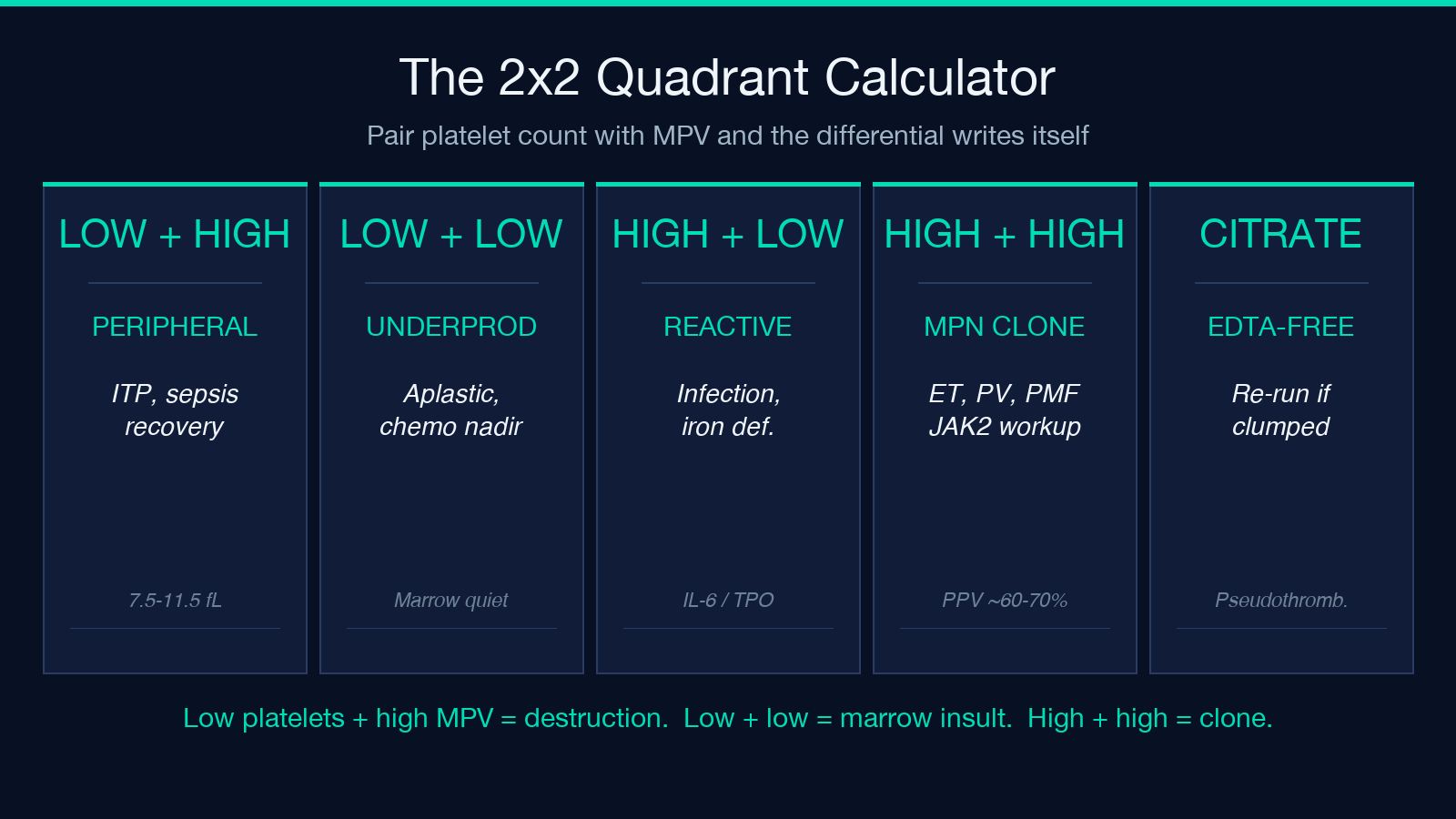
Low platelets plus high MPV is the classic pattern for peripheral destruction with compensatory turnover. The marrow is making platelets fine, but something is destroying them faster than the spleen can clear the debris. Immune thrombocytopenia (ITP) is the textbook example. Post-splenectomy patients also sit here because their platelet lifespan jumps from 7 to 10 days to nearly the full 10 days. Severe sepsis in recovery phase shows the same pattern as platelets rebound. Bernard-Soulier syndrome and MYH9-related disease are congenital macrothrombocytopenias that present in this quadrant from birth.
Low platelets plus low MPV indicates marrow underproduction. Aplastic anemia, recent chemotherapy or radiation, post-transfusion suppression, and iron-deficiency regeneration all land here. Wiskott-Aldrich syndrome, with its characteristically small platelets, also sits in this quadrant. The clinical implication is the opposite of the first quadrant: rather than suppressing immune destruction, you are looking for a marrow insult and treating it.
High platelets plus low MPV is reactive thrombocytosis. The marrow is responding to a cytokine signal (IL-6, TPO) but is not making young large platelets. Infection, post-surgical state, iron deficiency, and chronic inflammation all fit here. The platelet count can exceed 1000 x 10^9/L without any underlying myeloproliferative disease.
High platelets plus high MPV flags myeloproliferative neoplasms. Essential thrombocythemia, polycythemia vera, primary myelofibrosis, and chronic myeloid leukemia all present here. The combination of an elevated count and elevated MPV has a positive predictive value of 60 to 70 percent for a clonal process when the count is above 450 x 10^9/L, which is why most hematologists will order a JAK2 V617F mutation screen for this quadrant.
Where the Boundaries Get Blurry
Real patients do not always sit cleanly in one quadrant. Iron deficiency can shift from reactive thrombocytosis (high platelets, low MPV) toward a more ambiguous profile as iron stores recover, because newly synthesized platelets start larger. ITP patients on corticosteroids often move from low platelets + high MPV toward normal as the count recovers. Post-chemotherapy patients can land in low platelets + low MPV during the nadir and then sweep through all four quadrants over the recovery week.
There are also patients whose MPV is normal but whose platelet distribution width (PDW) is elevated. PDW is not on every analyzer, but when it is available, an elevated PDW with a normal MPV often means a mixed population of small and large platelets, which can be an early sign of recovery or an early sign of a clonal process. The MPV Analyzer works in MPV units specifically; if your analyzer outputs PDW instead, you need a different reference frame.
The Post-MI Cardioprotective Signal
Outside hematology, MPV has one of the strongest non-traditional prognostic signals in acute MI. Platelets in the hours after a coronary event are larger, more reactive, and contain more granular content. An MPV above 10.5 fL at admission correlates with higher 30-day mortality, larger infarct size, and worse reperfusion outcomes even after adjusting for GRACE and TIMI scores. The mechanism is platelet activation, not just acute-phase reaction. Patients with high MPV at presentation often need more aggressive antiplatelet loading because standard aspirin and P2Y12 doses underperform in this population.
This is one of the few clinical contexts where MPV is used for risk stratification rather than diagnosis, and the calculator does not address it directly. If you are building a post-MI risk model, you can wire MPV into the existing TIMI or GRACE calculators as a continuous variable.
Pre-Analytical Discipline Matters More Than the Tool
A calculator cannot save a sample that has been mishandled. Three rules cover the most common pre-analytical errors.
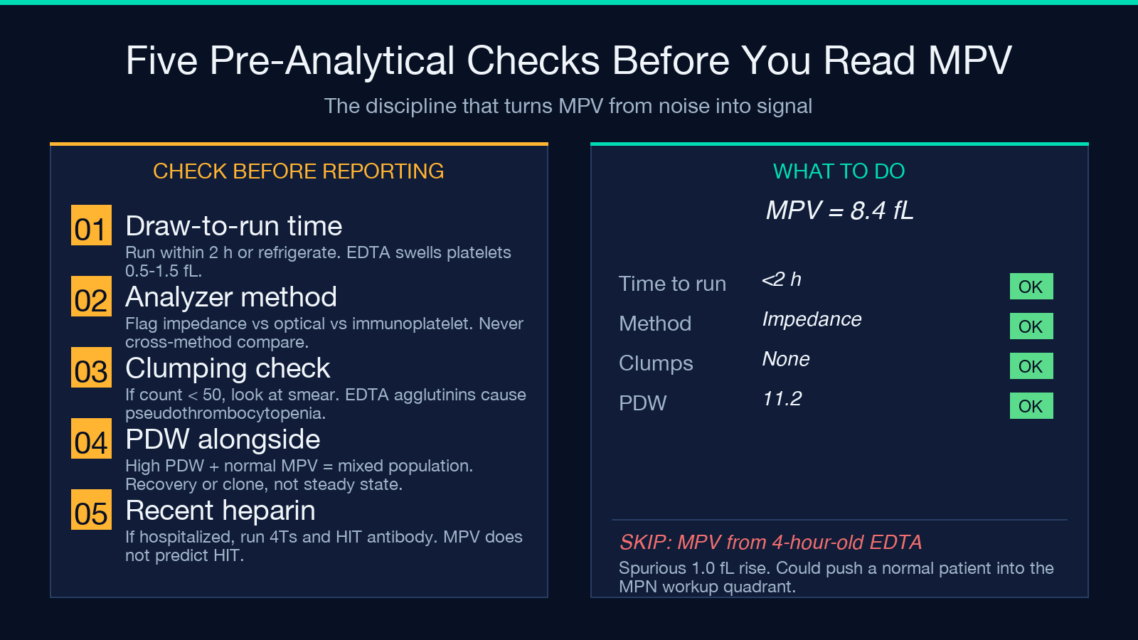
First, run the sample within two hours of draw or refrigerate it. EDTA swelling is real and predictable. Second, flag the analyzer method on every result so the clinician does not compare impedance MPV against optical MPV. Third, pair the MPV with the smear. A large platelet population on the smear without a corresponding MPV spike suggests platelet clumping, which is the most common pseudothrombocytopenia and is caused by EDTA-dependent agglutinins. The platelet count will read low (often below 50 x 10^9/L) but the patient has no bleeding risk because the clumped platelets are still functional. The MPV reading is unreliable in clumped samples; collect a citrate tube and rerun.
When to Trust the Quadrant, When to Order More Tests
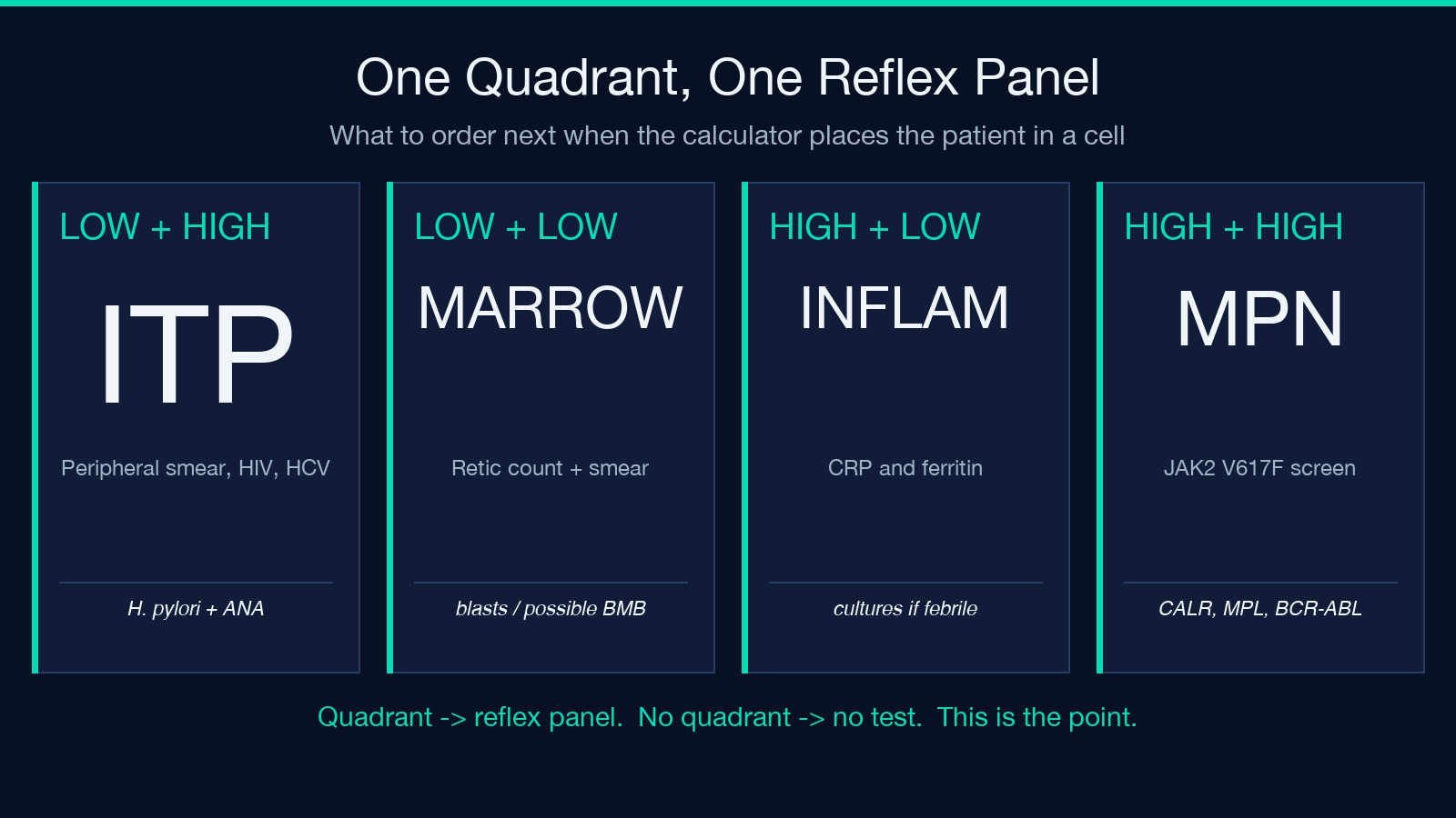
The 2×2 logic is a triage tool, not a diagnosis. Each quadrant corresponds to a different reflex test panel. Low platelets plus high MPV points to ITP workup (peripheral smear, HIV, HCV, H. pylori, ANA). Low platelets plus low MPV points to marrow assessment (reticulocyte count, peripheral smear for blasts, possibly bone marrow biopsy). High platelets plus low MPV points to inflammatory or infectious workup (CRP, ferritin, blood cultures if febrile). High platelets plus high MPV points to MPN workup (JAK2 V617F, CALR, MPL, BCR-ABL).
If the patient has been on heparin in the last two weeks, add a HIT antibody screen regardless of quadrant. Heparin-induced thrombocytopenia does not fit cleanly into the differential because the antibody targets platelet factor 4 and triggers consumption through a mechanism independent of platelet size. The 4Ts score should run in parallel.
Using the Calculator Effectively
The Mean Platelet Volume Analyzer is a two-input tool. Enter MPV in fL and the platelet count in 10^9/L. The output is the quadrant plus a short list of conditions that fit. The tool does not store patient data and does not require an account, which makes it usable at the bedside or during chart review. If you want to extend it with PDW, reticulated platelet percentage, or immature platelet fraction, those are not on the tool today but the 2×2 framework extends naturally.
For laboratory directors building the differential logic into an LIS rule, the same four-quadrant framework can be encoded as a reflex test suggestion. Low platelets plus high MPV with a known ITP history should suppress the ITP workup reflex and instead trigger a heparin antibody screen if the patient has been hospitalized. This kind of contextual rule is exactly the kind of decision support that MPV has historically lacked in standard EMR CBC result displays.
What MPV Does Not Tell You
MPV is a size measurement, not a function measurement. A large platelet can still be dysfunctional, and a small platelet can still be hyperreactive. Platelet function testing (light transmission aggregometry, VerifyNow, PFA-100) addresses activation, not size. Do not substitute MPV for a functional assay when the question is whether aspirin is working or whether a patient with a bleeding history has a storage pool defect.
Also, MPV is not a screening test for bleeding risk in unselected patients. The MPV distribution in healthy adults overlaps substantially with the distribution in patients with mild platelet function disorders. A normal MPV does not rule out a storage pool defect, and an elevated MPV does not predict bleeding. Use MPV where the evidence base supports it: thrombocytopenia differential, post-MI risk, and MPN screening in patients with elevated counts. Outside those contexts, treat MPV as exploratory.
The cheapest signal on a routine CBC is also one of the most underused. Run the tool on the next thrombocytopenia you see, and the quadrant will tell you where to look before you order the next test. Explore more CBC and hematology tools at Elysia Tools.
