Every biology student has stared at a long DNA string and tried to slide a window three letters at a time, hunting for the next “ATG.” The window is the trick. Three letters, no commas, no spaces, no pause until the ribosome reaches a stop. The 64-cell codon table is what every window reads against. The translator’s whole job is to slide that window across a sequence in all three forward frames, then build the reverse complement and slide again across three more frames — six in total — and hand back every amino acid chain it can find. The interesting part is not the lookup. The interesting part is what the six frames reveal that one frame hides.
This is a walkthrough of the Codon to Amino Acid Translator at Elysia Tools: what a reading frame is, why six frames are necessary, where the standard genetic code stops and the mitochondria take over, and when a tool like this actually saves your afternoon versus when you should reach for a full ORF finder. If you have a candidate coding sequence in hand and want the amino acid chain in under a second, the translator is the shortest path; everything below is the mental model behind what the tool is actually doing when you click “translate.”
The Codon Table Is Smaller Than You Think
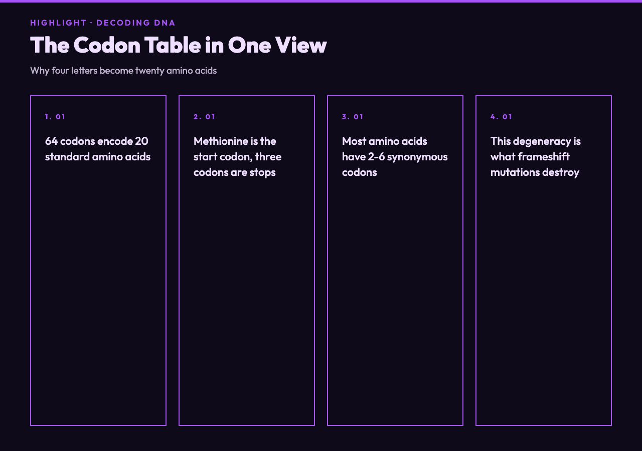
There are four nucleotide letters — A, C, G, T in DNA; A, C, G, U in mRNA — and you read them three at a time. That gives 4³ = 64 possible codons. Sixty-one of those code for the twenty standard amino acids. Three do not code for anything: TAA, TAG, and TGA. Those are the stop codons, and a translator’s first job is to recognize them.
Methionine is encoded by ATG and, almost always, ATG is also the start codon. That single fact is what makes reading frames tractable. A long DNA strand can have ATG sitting in many positions, but only one of those positions is in the correct frame for the actual gene. Without a start marker, you would not know which frame is the “right” one; with ATG, you get an anchor. The translator marks every ATG it finds, labels it Met, and continues reading the downstream codons in that frame until it hits a stop. Each such run is an open reading frame — an ORF.
Notice that four amino acids are encoded by six codons each (leucine, serine, arginine), five are encoded by four codons, nine by two, and two — methionine and tryptophan — by exactly one. That degeneracy is why frameshift mutations are so destructive: a single insertion or deletion shifts every codon downstream, so the amino acid sequence changes even when only one nucleotide moved. The translator makes that visible by labeling every codon with its amino acid; run a wild-type and a single-base-insertion mutant through it and the resulting diff is impossible to misread.
Why Six Frames and Not One
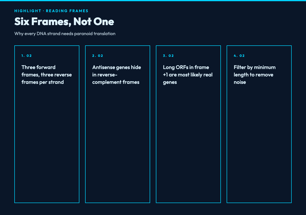
Given a 1,000-nucleotide strand, where is the gene? You cannot know by inspection. The cell knows because the ribosome binds at a specific mRNA position and reads from there — but you, looking at raw sequence, do not have that signal. So you check every possible frame. Three forward frames start at nucleotide 1, 2, and 3 of the strand. Three reverse frames start at nucleotide 1, 2, and 3 of the reverse complement. Every gene in a sequenced genome lives in one of those six, and the translator scans all of them.
The practical reason this matters: upstream annotation can be wrong, or the gene you care about can be on the antisense strand. If you only scan frame +1, you will miss every antisense ORF, every gene with a leader peptide upstream of the canonical start, and every retroviral gene that the host reads in an unexpected direction. The six-frame approach is paranoid by design — it produces more candidate ORFs than biology will use, and the user filters down.
The translator’s output is typically a list of all ORFs above a configurable minimum length, grouped by frame, with the amino acid sequence written in one-letter code. Long ORFs in frame +1, +2, or +3 of the forward strand are most likely to be real. Anything in a reverse-complement frame is a hint to look at the opposite strand of your input. The translator does all of this in a single click — paste, choose the translation table, hit translate — and you get the six frames back as plain text you can diff against your reference protein.
Start Codons, Stop Codons, and the Edge Cases That Catch People
Most bacterial genes start with ATG and end with TAA, TAG, or TGA. Most eukaryotic mRNAs do the same, but with a twist: the start codon is recognized in a Kozak context (gccRccAUGG, where the capital A is the +1 nucleotide of ATG), so not every ATG is actually a start site in vivo. The translator does not know about Kozak context — it treats every ATG as a start candidate and every in-frame TAA/TAG/TGA as a stop. That is the right behavior for sequence inspection: the user gets all candidates, then layers biological knowledge on top.
Stop codons are not always stops. In some contexts TGA codes for selenocysteine (the 21st amino acid), and TAG occasionally codes for pyrrolysine (the 22nd). Mitochondria use a slightly different code: AGA and AGG are stops in vertebrate mitochondria but code for arginine in the standard code; ATA codes for methionine in mitochondria but for isoleucine in the standard code; and TGA codes for tryptophan in mitochondria, not stop. The translator lets you switch between the standard, vertebrate mitochondrial, yeast mitochondrial, and a few other NCBI translation tables. Pick the wrong one and every ORF in a mitochondrial gene will appear garbled.
Single nucleotide changes inside a codon produce one of three outcomes: a synonymous substitution (same amino acid, because of codon degeneracy), a missense substitution (different amino acid), or a nonsense substitution (a stop codon appears where an amino acid used to be). The translator does not classify these for you — it just emits the amino acid chain — but if you translate the wild-type and the mutant and diff the two outputs, every change you see is a real amino acid change, and every position where the chain shortens is a nonsense mutation.
Frameshifts, the Translator’s Stress Test
Insert or delete a single nucleotide and every codon downstream shifts by one. The result is a downstream amino acid sequence with no relationship to the original — and, usually, a premature stop codon that truncates the protein. This is why indel mutations are so much more damaging than point mutations. The translator makes frameshifts visible: translate the wild-type, translate the mutant, the difference is impossible to miss.
If you want to debug a suspicious frameshift, the workflow is straightforward. Translate both strands of both alleles in all six frames. The frame that produces a clean, full-length ORF in the wild-type is your gene. Translate the mutant in the same frame and look for the position where the amino acid chain diverges. That position is one nucleotide downstream of the actual indel.
There is also the rarer case of programmed ribosomal frameshifting, where the ribosome intentionally slips back by one nucleotide (−1) or forward by one (+1) at a specific sequence signal. This happens in some retroviruses and in a handful of cellular genes. The translator cannot detect these — by design, it reads each frame rigidly — but it is worth knowing the limitation exists before assuming a frameshift is a mutation.
When the Translator Is Enough, and When You Need More
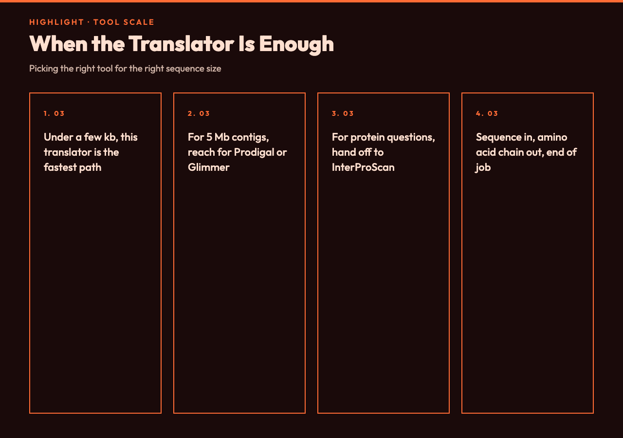
The translator is the right tool when you have a single ORF in mind, a candidate coding sequence under a few kilobases, or a clean wild-type/mutant comparison. It is fast, deterministic, and gives you the answer in amino acid letters without intermediate files. Six frames, all six translation tables, copy-to-clipboard output — that’s the bread and butter.
It is the wrong tool when you have a 5 Mb contig and no annotation. For that scale, you want a real ORF predictor: Prodigal, Glimmer, or MetaGeneAnnotator, which combine codon statistics, GC content, and Shine-Dalgarno motif detection to rank candidate genes. Those tools can process a bacterial genome in seconds and return hundreds of ranked ORFs with confidence scores. The translator does not rank — it enumerates — so for a large input you would drown in false-positive short ORFs (every random ATG produces a 3-codon ORF, which is noise).
It is also the wrong tool for protein-level questions: secondary structure, domain annotation, signal peptide prediction, transmembrane topology. Once you have the amino acid sequence, hand it to a domain predictor like InterProScan or a structure predictor like AlphaFold. The translator’s job ends where the protein begins. For everything up to that boundary — sequence in, amino acid chain out — the Codon to Amino Acid Translator is the lightest-weight tool in your browser.
A Few Sibling Tools Worth Pairing With This One
If you do this often, a few Elysia Tools live next door and handle the upstream and downstream work. The Codon to Amino Acid Translator is the centerpiece, but the rest of the workflow is just as clickable.
- Reverse complement — needed before you can scan the antisense strand. Most translators, including this one, do it for you, but if you want to inspect the complement separately the tool is one click away.
- GC content calculator — useful for sanity-checking codon usage bias. A gene with very high GC content will have a heavily skewed codon table even within the same amino acid.
- FASTA / sequence formatters — most public databases (NCBI, Ensembl, ENA) deliver sequences wrapped at 60 or 80 characters; you want them unwrapped before you paste them into the translator.
- Hash / checksum tools — handy for tagging a sequence with a stable identifier so you can compare two translated outputs across sessions.
- JSON formatter — the translator’s structured output, when you copy it as JSON, sometimes lands minified; one click returns it readable.
- Markdown to HTML — when you are pasting translation output into a lab notebook or a blog post and want the amino acid chain to render cleanly.
- Character / string utilities — for stripping whitespace, normalizing line endings, or removing FASTA headers before you paste.
That is the surrounding toolset — none of them is a substitute for the translator, but they all sit one click away on the same site and remove the small friction that accumulates when you are doing this for the twentieth time in an afternoon.
Putting It Together
A DNA strand is a one-dimensional string. A protein is a one-dimensional string. The translator is the deterministic function between them, parameterized by which frame you start at and which genetic code you use. Six frames, four translation tables (in practice), and you have enumerated every possible protein the strand could encode. Most of those are noise — short ORFs, antisense collisions, sequences outside the actual gene — but the one that survives your minimum-length filter and your biological sanity check is your answer. The tool does not think. It enumerates. That is exactly what you want when you are debugging a mutation, checking an ORF you found by BLAST, or verifying a gene synthesis before you order it.
The Codon to Amino Acid Translator lives at elysiatools.com, free, no signup, paste-and-go. The first time you use it on a wild-type/mutant pair and watch a 6-line amino acid diff appear, the value is obvious. The second time, you wonder how you ever debugged ORF frameshifts without it.