
A thyroid panel does not give a diagnosis. It gives a three-number pattern, and the diagnosis lives in the pattern. Two patients with the same TSH can sit on opposite sides of the thyroid spectrum if their FT4 moves in opposite directions; a third patient can look identical on TSH and FT4 yet still need a different workup because FT3 is the tipping number. The Thyroid Index tool reads the panel the way an endocrinologist reads it — by first classifying the TSH-FT4 axis, then layering FT3 on top, then flagging the patterns that change management. This field guide walks through every branch of that decision tree, with the unit conversions, edge cases, and follow-up logic that turn a single panel into a working differential.
What “TSH” Actually Measures and Why It Moves
TSH is the pituitary’s read of thyroid hormone availability at the tissue level, not a direct read of the gland. The hypothalamus secretes TRH, the anterior pituitary secretes TSH in response, TSH drives the thyroid to produce T4 (mostly) and a small amount of T3, and both T4 and T3 feedback negatively on the pituitary. A high TSH means the pituitary is calling for more hormone because circulating T4 and T3 are inadequate; a low TSH means the pituitary is being told to stand down because circulating hormone is high. That feedback loop is log-linear — a doubling of free T4 typically suppresses TSH by an order of magnitude — which is why a tiny FT4 shift can produce a large TSH shift while the patient still feels well.
Two operational consequences follow. First, TSH on its own is never the answer; the FT4 must be in the same report or the panel is uninterpretable. Second, TSH is suppressed by a long list of non-thyroidal inputs — dopamine, glucocorticoids, severe illness, cabergoline, even pregnancy in the first trimester (beta-hCG cross-reacts with the TSH receptor). When the picture does not fit, ask what else is talking to the pituitary before you ask what the thyroid is doing.
The reference ranges the Thyroid Index uses for adults are: TSH 0.4–4.0 mIU/L (or µIU/mL — numerically identical), FT4 0.8–1.8 ng/dL equivalent to 10–23 pmol/L, FT3 2.3–4.2 pg/mL equivalent to 3.5–6.5 pmol/L. Trimester-specific pregnancy ranges, pediatric ranges, and the post-thyroidectomy suppressed range are narrower; the tool accepts a units toggle for each hormone so a European lab reporting FT4 in pmol/L lands in the same reference band as a US lab reporting ng/dL.
The Six Pattern Branches
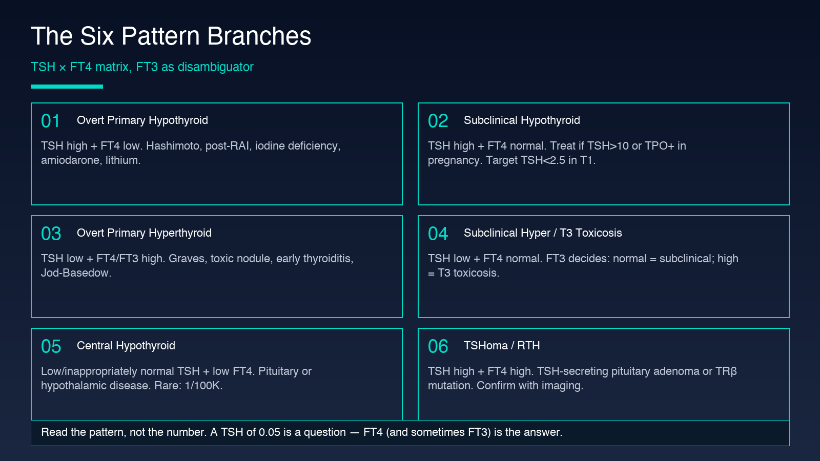
Once you have TSH and FT4, the diagnostic tree is a 2×3 matrix. TSH is high, normal, or low; FT4 is high, normal, or low. Each of the six cells has a canonical interpretation, and the FT3 is the disambiguator for the cells where the answer depends on the T4-to-T3 conversion axis. The next three sections walk through the high-TSH, low-TSH, and rare-cell branches in clinical detail.
High TSH, Low or Normal FT4 — Hypothyroid Spectrum
High TSH + Low FT4 is the textbook Hashimoto’s cell. The thyroid is failing (low FT4) and the pituitary is shouting (high TSH). Common causes: Hashimoto thyroiditis (anti-TPO and anti-thyroglobulin positive in 80 percent of cases), post-ablative hypothyroidism after radioiodine or thyroidectomy, iodine deficiency in endemic regions, and drug-induced suppression (amiodarone, lithium, interferon-alpha, immune checkpoint inhibitors). The clinical picture is fatigue, cold intolerance, constipation, dry skin, weight gain, and a slow deep-tendon-reflex relaxation phase. Treatment is levothyroxine replacement, typically 1.6 µg/kg lean body weight per day, with TSH rechecked at 6–8 weeks. The Thyroid Index flags this combination as overt primary hypothyroidism and surfaces the Hashimoto/post-ablation/iodine/amiodarone list as the common cause panel.
High TSH + Normal FT4 is subclinical hypothyroidism — defined by an elevated TSH with a normal FT4 in an asymptomatic or minimally symptomatic patient. About 2–5 percent of adults sit here; the prevalence is higher in women over 60 and in iodine-replete populations. The decision to treat depends on TSH level, antibody status, and clinical context. A TSH above 10 mIU/L generally warrants treatment regardless of antibodies. A TSH between 4.5 and 10 in a TPO-antibody-positive patient is often treated; the same range in a TPO-antibody-negative patient is often watched. Pregnancy is a hard indication to treat — even subclinical hypothyroidism is associated with miscarriage and preterm birth, and the target TSH in pregnancy is below 2.5 in the first trimester and below 3.0 in later trimesters.
Low TSH, High or Normal FT4 — Hyperthyroid Spectrum
Low TSH + High FT4 (or High FT3) is overt primary hyperthyroidism. The thyroid is overproducing, FT4 is above range, and TSH is suppressed. The differential is Graves disease (TSH-receptor antibodies positive, often with ophthalmopathy and pretibial myxedema), toxic multinodular goiter, toxic adenoma, and the early phase of subacute or painless thyroiditis. Iodine-induced hyperthyroidism (Jod-Basedow) is a classic trigger in patients given iodinated contrast or started on amiodarone in the setting of underlying nodular autonomy. The clinical picture is the opposite of hypothyroidism: heat intolerance, weight loss, palpitations, tremor, diarrhea, hyperreflexia. Treatment depends on the cause — antithyroid drugs (methimazole, PTU) for Graves, surgery or radioiodine for toxic nodules, beta-blockers for symptom control across all causes.
Low TSH + Normal FT4 is the cell where FT3 changes the answer. This is subclinical hyperthyroidism if FT3 is also normal — usually exogenous thyroid hormone over-replacement in a hypothyroid patient, autonomous thyroid nodules producing just enough to suppress TSH without spilling FT4 above range, or resolving thyroiditis where the gland has begun to recover from an inflammatory destructive phase. The same low-TSH-normal-FT4 cell becomes T3 toxicosis if FT3 is elevated — some autonomous nodules and some Graves variants preferentially secrete T3 over T4, and the diagnosis flips to overt hyperthyroidism. The Thyroid Index prompts for FT3 in this branch specifically because the answer hinges on it. A low TSH with normal FT4 in a sick hospitalized patient is also a common pattern — the non-thyroidal illness syndrome, which is usually not treated and resolves with the underlying illness.
Rare Cells: Central Hypothyroid and TSHoma
Low or Normal TSH + Low FT4 is central hypothyroidism — either secondary (pituitary disease) or tertiary (hypothalamic disease). The TSH is misleadingly low or inappropriately normal because the feedback loop is broken at the regulator. This is rare — about 1 in 100,000 — but it is easy to miss because the pattern looks like mild hypothyroidism at first glance. The clinical clue is that the TSH is not high when FT4 is low. Workup includes a full pituitary panel (cortisol, LH, FSH, prolactin, IGF-1) and pituitary MRI. Treatment is levothyroxine replacement, but the target is FT4 in the upper half of normal, not TSH normalization, because the TSH is unreliable.
High TSH + High FT4 should not exist in a normal feedback loop. Two possibilities: a TSH-secreting pituitary adenoma (TSHoma) producing TSH that is bioactively elevated and not properly suppressed, or thyroid hormone resistance (RTH), most often from a mutation in the thyroid hormone receptor beta (TRβ) gene. The distinction is usually made with an alpha-subunit measurement, sex hormone-binding globulin (SHBG), and pituitary MRI; RTH has a normal pituitary and a family history, while TSHoma has an elevated alpha-subunit ratio and a pituitary mass. Treatment differs — TSHoma is usually resected; RTH is usually observed unless symptomatic, in which case beta-blockers or triiodothyroacetic acid may help.
The Low-T3 Syndrome Pattern
A separate pattern deserves its own callout: low FT3 with normal TSH and normal FT4. This is the non-thyroidal illness syndrome or low-T3 syndrome, seen in severe systemic illness, fasting, post-operative states, and chronic inflammatory conditions. The mechanism is increased peripheral conversion of T4 to reverse T3 (rT3) at the expense of T3. Most experts do not recommend treating this pattern with thyroid hormone; the consensus is that the thyroid axis is responding appropriately to systemic stress and that the low T3 is a marker rather than a cause. The exception is in chronic kidney disease and some chronic heart failure populations, where low T3 is prognostic and small trials of T3 replacement have shown benefit, though this remains investigational.
The Caveats That Break the Pattern
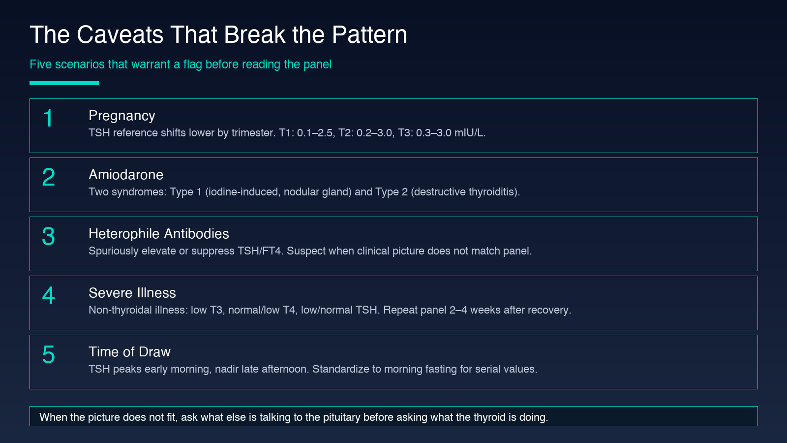
Five clinical scenarios break the clean branching logic above and warrant flagging in the Thyroid Index interpretation. Pregnancy suppresses TSH in the first trimester via beta-hCG, so the normal reference shifts lower; use trimester-specific ranges (T1: 0.1–2.5, T2: 0.2–3.0, T3: 0.3–3.0 mIU/L). Amiodarone contains a large iodine load and blocks T4-to-T3 conversion, causing two distinct thyroid syndromes: type 1 (iodine-induced hyperthyroidism in a nodular gland) and type 2 (destructive thyroiditis). Heterophile antibodies can spuriously elevate or suppress TSH/FT4 depending on the platform — suspect when the clinical picture does not match the panel. Severe illness produces the non-thyroidal illness pattern (low T3, normal or low T4, normal or low TSH) that can mimic central hypothyroidism; repeat the panel 2–4 weeks after recovery before making a new diagnosis. And TSH has diurnal variation with a peak in early morning and a nadir in late afternoon, so standardize the draw to morning fasting when comparing serial values in the same patient.
How to Use the Tool in a 30-Second Read
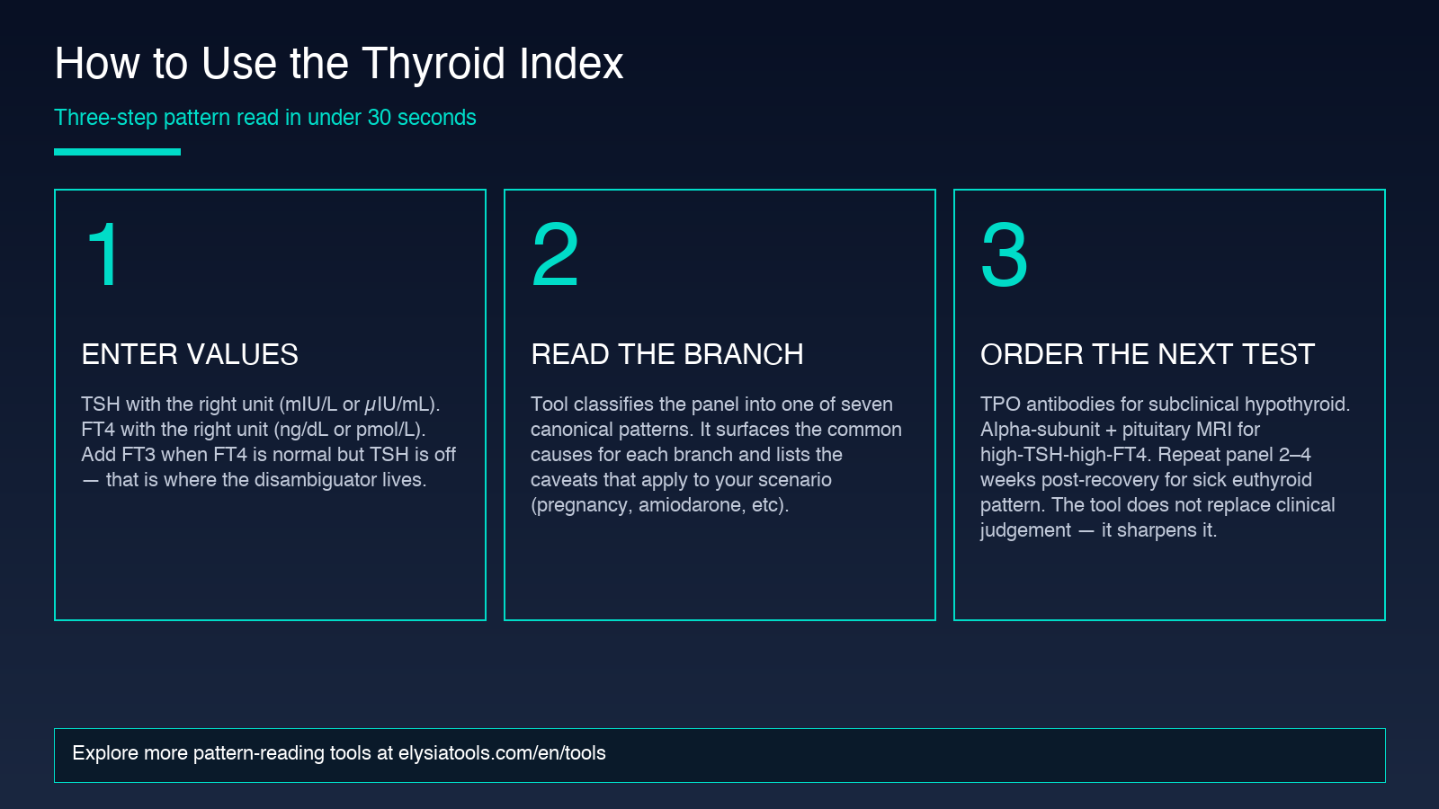
The Thyroid Index is built to compress the pattern-recognition work above into three quick steps. Enter TSH with the correct unit, enter FT4 with the correct unit, and (when the result requires it) enter FT3. The tool classifies the pattern into one of the canonical branches above, returns the canonical interpretation, lists the common causes, and surfaces the caveats that apply. It does not replace clinical judgement — the disclaimer in the tool’s footer is explicit about that — but it makes the pattern-to-diagnosis mapping fast and consistent, and it catches the disambiguating cases where FT3 or further workup changes the answer. For trainees, it is a useful way to learn the branching logic. For clinicians, it is a sanity check before ordering the next test or starting treatment.
The single most important habit the tool reinforces: read the pattern, not the number. A TSH of 0.05 is not a diagnosis — it is a question. The diagnosis is in the FT4, and sometimes in the FT3, and always in the clinical context.
Explore more pattern-reading tools at elysiatools.com.
